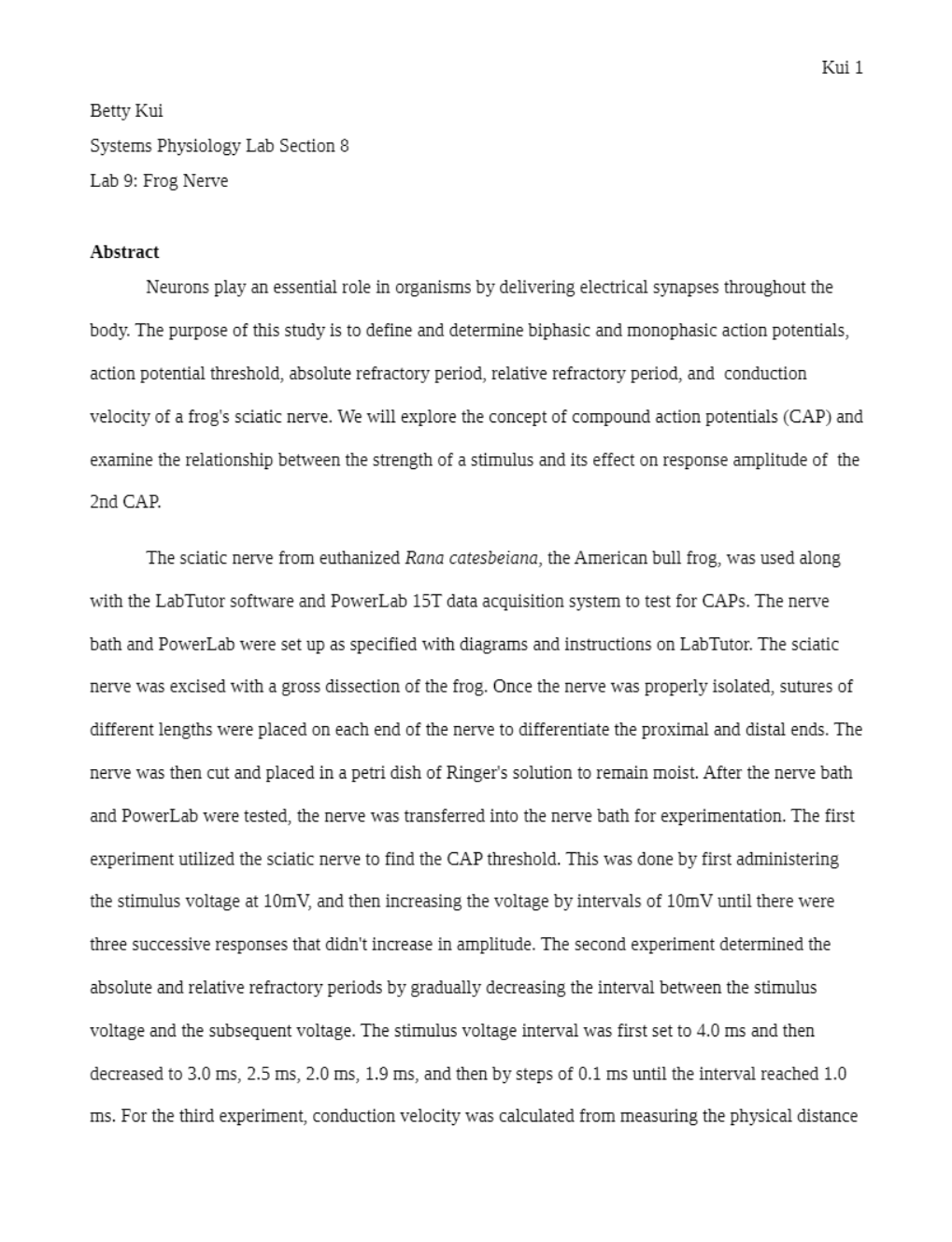
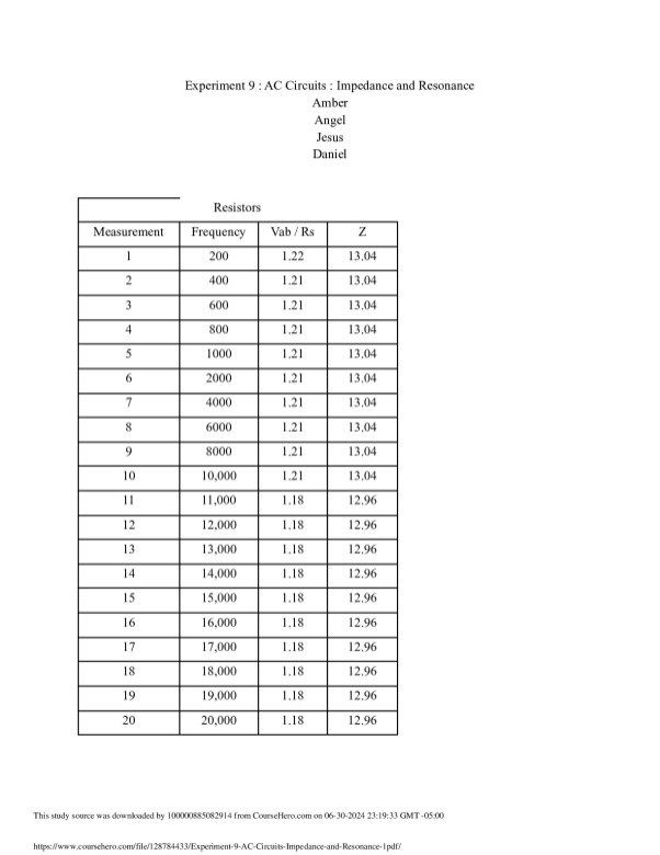
AbstractNeurons play an essential role in organisms by delivering electrical synapses throughout thebody. The purpose of this study is to define and determine biphasic and monophasic action potentials,action potential threshold, absolute refractory period, relative refractory period, and conductionvelocity of a frog's sciatic nerve. We will explore the concept of compound action potentials (CAP) a
...[Show More]
Abstract
Neurons play an essential role in organisms by delivering electrical synapses throughout the
body. The purpose of this study is to define and determine biphasic and monophasic action potentials,
action potential threshold, absolute refractory period, relative refractory period, and conduction
velocity of a frog's sciatic nerve. We will explore the concept of compound action potentials (CAP) and
examine the relationship between the strength of a stimulus and its effect on response amplitude of the
2nd CAP.
The sciatic nerve from euthanized Rana catesbeiana, the American bull frog, was used along
with the LabTutor software and PowerLab 15T data acquisition system to test for CAPs. The nerve
bath and PowerLab were set up as specified with diagrams and instructions on LabTutor. The sciatic
nerve was excised with a gross dissection of the frog. Once the nerve was properly isolated, sutures of
different lengths were placed on each end of the nerve to differentiate the proximal and distal ends. The
nerve was then cut and placed in a petri dish of Ringer's solution to remain moist. After the nerve bath
and PowerLab were tested, the nerve was transferred into the nerve bath for experimentation. The first
experiment utilized the sciatic nerve to find the CAP threshold. This was done by first administering
the stimulus voltage at 10mV, and then increasing the voltage by intervals of 10mV until there were
three successive responses that didn't increase in amplitude. The second experiment determined the
absolute and relative refractory periods by gradually decreasing the interval between the stimulus
voltage and the subsequent voltage. The stimulus voltage interval was first set to 4.0 ms and then
decreased to 3.0 ms, 2.5 ms, 2.0 ms, 1.9 ms, and then by steps of 0.1 ms until the interval reached 1.0
ms. For the third experiment, conduction velocity was calculated from measuring the physical distance
This study source was downloaded by 100000793680026 from CourseHero.com on 04-20-2021 18:29:29 GMT -05:00
https://www.coursehero.com/file/19718865/Systems-Frog-Nerve-Lab/
This study resource was
shared via CourseHero.com
Kui 2
between the recording electrodes and finding distance between the nearest and further CAPs in the
waveform on LabTutor. The experiments yielded that an increase in the strength of a stimulus voltage
will initially increase the CAP amplitude until 70 mV but will decrease afterwards and plateau at
higher voltages. Data also suggest a trend that as stimulus interval decreased, 2nd CAP amplitude
decreased. 1.9 ms marked the end of the absolute refractory period and beginning of the relative
refractory period for the sciatic nerve. Conduction velocity was calculated to be less than normal
values found in other studies which could be explained by Ringer's solution possibly coming in contact
with the wires shorting the stimulus electrode. These finding illustrate the potential for further studies
to be performed to expand knowledge about action potential firing within organisms.
Introduction
Action potentials are brief fluctuations in membrane potential which enable a method of
communication between nerves to maintain life in organisms. Action potentials are a caused by the
rapid opening and closing of voltage-gated ion channels. First, an influx of Na+ into the cell causes
depolarization. As more and more Na+ ions migrate, the potential rises to about +35 mV causing Na+
channels to close (Brink, 1983). K+ channels open allowing K+ to leave repolarizing the membrane
(Brink, 1983). However, the potassium pump oftentimes does not close quick enough causing an
overshoot known as hyperpolarization which makes a brief drop past the resting potential (Brink,
1983). This illustrates how most models of cell electrical activity show that only the motion of positive
ions, and specially those of potassium, calcium and sodium, influence membrane potential (Endresen,
2000). This membrane potential can be measured by calculating the potential difference between two
electrodes placed on the surface of a single nerve. A nerve is a collection of axons of many neurons
and these axons have varying thicknesses that would affect the speed and size of their action potentials
(Intermediate Physiology Handbook, 2004). The action potentials, recorded from the outside of the
nerve is known as a compound action potential (CAP) and represents the sum of all the action
This study source was downloaded by 100000793680026 from CourseHero.com on 04-20-2021 18:29:29 GMT -05:00
https://www.coursehero.com/file/19718865/Systems-Frog-Nerve-Lab/
This study resource was
shared via Co
[Show Less]


-preview.png)